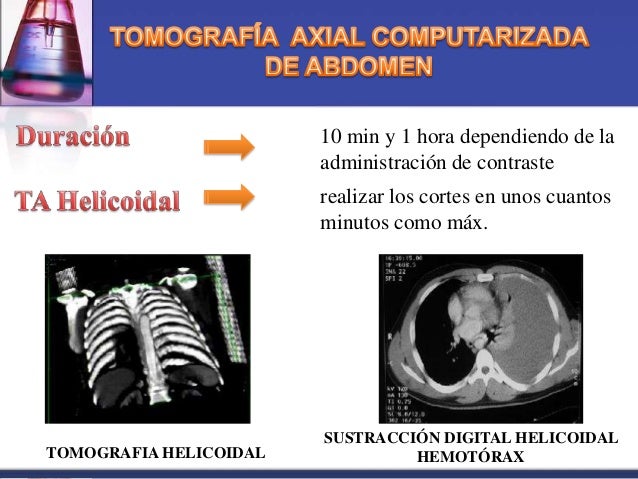

First phase was a 60 mL bolus of iopramide IV, at a rate of 2.0 mL/s in 30 seconds, followed by an iopromide administration pause of 45 seconds. A total of 130 mL of contrast, with biphasic injection technique, was used with an inter-bolus delay of 45 seconds. Whippany, NJ: Bayer Health Care Pharmaceuticals) was administered via an 18-gauge peripheral intravenous (IV) catheter. Low osmolar non-ionic contrast medium (Iopromide Ultravist R. Whole-body computed tomography included a non-contrast brain and a contrasted neck, chest, abdomen and pelvis (from the base of the skull to lower edge of the pubis) as a single-pass computed tomography helical acquisition ( Table 1). Resuscitation was initiated in the trauma bay and continued during scanning. The radiologist read each study in real time. Each patient was accompanied by the trauma team (Trauma and Acute Care Surgeon, Fellow, Surgery Resident, Emergency Physician and Trauma Nurses). The scanner is located adjacent to the trauma bay (<100 feet).
Tomografia computarizada sin contraste software#
Calculated survival rates were assessed using the Trauma and Injury Severity Score (TRISS), which uses clinical parameters to determine the probability of survival, in order to perform comparisons with the real survival rate and evaluate possible impact on mortality with our management algorithm.ĭata was acquired using a multi-slice IVR- computed tomography system (Aquilion ONE 320 Slice computed tomography scanner, software version 7.0, Toshiba Medical Systems Corp., Tochigi, Japan). Data on demographics, trauma mechanism, clinical severity (Injury Severity Score (ISS), New Injury Severity Score (NISS), SOFA Scale) and outcomes (surgical or non-surgical management, ICU admission and mortality) were extracted from clinical records. Patients who were transferred from or to another institution were excluded, this way only patients with complete information and follow-up were included. All adult patients (>15-years) who met these criteria and underwent whole-body computed tomography between January, 2017 and December, 2018 were included and followed-up by a research assistant. The institutional single-pass whole-body computed tomography protocol of Fundación Valle del Lili (a Level I Trauma Center in Cali, Colombia) was created in 2016 establishing the indication for whole-body computed tomography in all patients with severe polytrauma: moderate to severe head trauma, suspected solid organ injury, hollow viscus or vascular injury, pelvic trauma and unstable fractures that arrived hemodynamically stable or unstable. With prior institutional ethics approval, a descriptive evaluation of patients who underwent single-pass whole-body computed tomography was performed. The objective of this study was to describe the implementation of a new institutional single-pass whole-body computed tomography protocol in the management of patients with severe blunt/penetrating trauma, hemodynamically stable or unstable, as a diagnostic tool for the decision making between surgical or non-surgical treatment.
Achieving an optimal equilibrium phase is important to visualize arterial and venous phases in a single image acquisition, to identify the hemorrhage source and reduce the time in the computed tomography suite. Many different whole-body computed tomography protocols have been used worldwide, but the optimal technique is still not clear However, its use has been limited in hemodynamically unstable trauma patients because of the potential delay in therapeutic interventions

In addition, compared with selective images, Whole-body computed tomography can identify less evident lesions that could go undetected, translating to improved prognosis, lower mortality and less waiting time in the emergency department Without a significative increase in overall costs Moreover, there is a growing body of evidence on the benefits of this diagnostic method, increasing the probability of survival in polytrauma patients Whole-body computed tomography is currently the standard method of workup recommended for the primary evaluation of trauma patients at many centers because of its high sensitivity and specificity for the diagnosis of injuries, in cases of abdominal trauma, it reaches a sensitivity of 94% and specificity of 95%, superior to ultrasound and clinical evaluation (S: 30-75% E: 30-50%) During initial evaluation of trauma patients in the emergency department, prompt attention by the trauma team, early diagnosis and effective treatment have been the main survival determinants Trauma is the second cause of death in men of all ages, accounting for 7% of all deaths in Colombia in 2018


 0 kommentar(er)
